
They say a picture is worth a thousand words. In NEET Biology, a picture might just be worth a seat in a government medical college.
With nearly 20–25% of NEET Biology questions being diagram-based or derived from labels, your ability to visualise the 'plant cell' or the 'structure of mitochondria' is your biggest competitive advantage.
Let’s dive into the essential visual checklist every NEET aspirant needs to master.
Most Important NEET Diagrams in NCERT Class 11 Biology
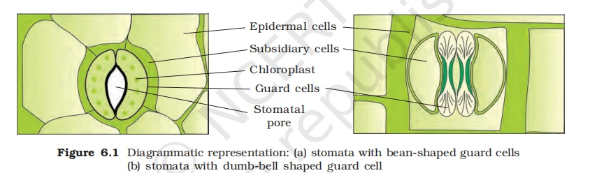
NEET Biology Diagrammatic representation of stomata
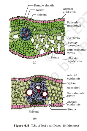
NEET Biology T.S Dicot and Monocot of leaf
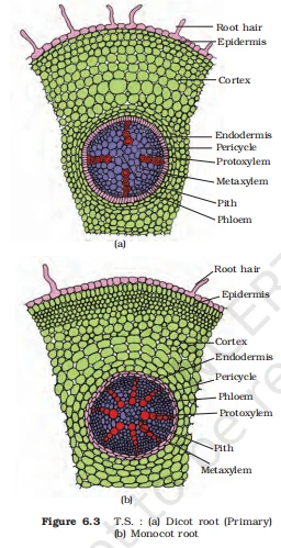
NEET Biology T.S Dicot and Monocot of root
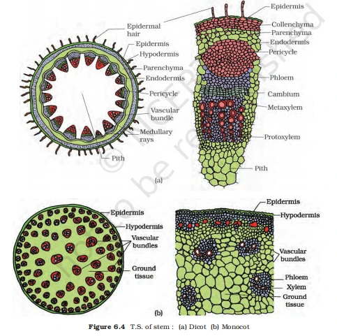
NEET Biology T.S Dicot and Monocot of stem
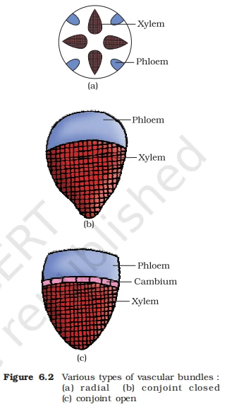
NEET Biology Various types of vascular bundles
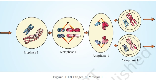
NEET Biology Stages of Meiosis 1 (Cell Cycle and Cell Division)
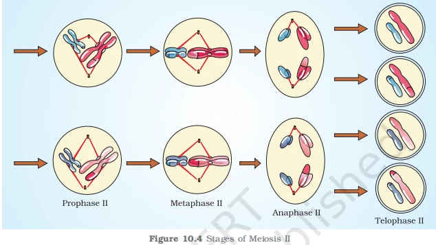
NEET Biology Stages of Meiosis 2 (Cell Cycle and Cell Division)
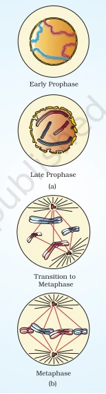
NEET Biology Stages of Mitosis (Cell Cycle and Cell Division)
NEET Biology Stages of Mitosis 2 (Cell Cycle and Cell Division)
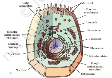
NEET Biology Animal Cell (Cell The Unit of Life)
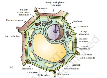
NEET Biology Plant Cell (Cell The Unit of Life)
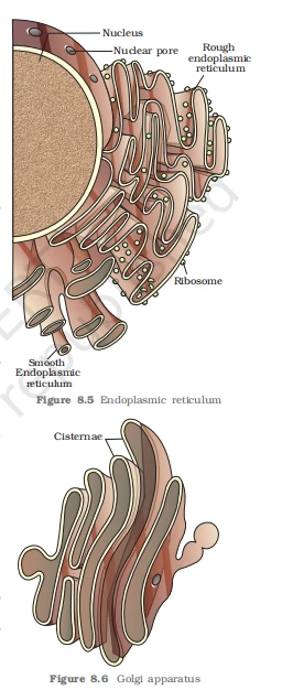
NEET Biology Endoplasmic Reticulum and Golgi apparatus
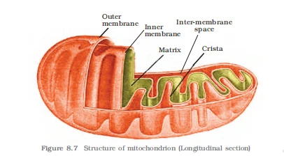
NEET Biology Structure of Mitochondria (Cell The Unit of Life)

NEET Biology Sectional View of Chloroplast (Cell The Unit of Life)
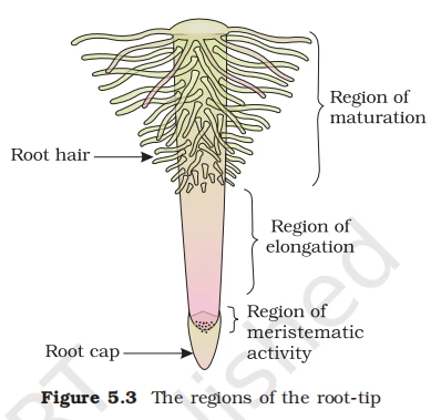
NEET Biology Regions of root-tip
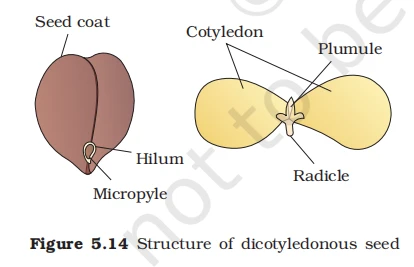
NEET Biology Structure of dicot
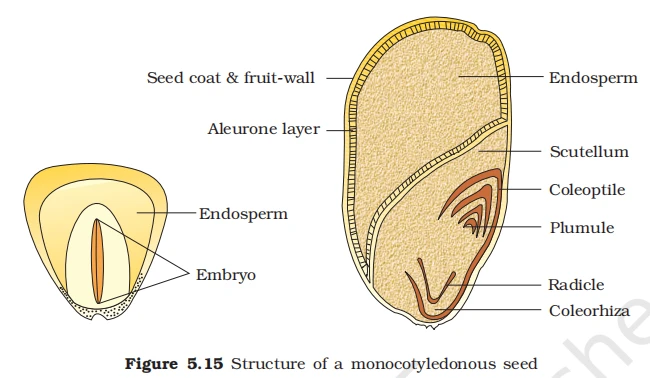
NEET Biology Structure of monocot
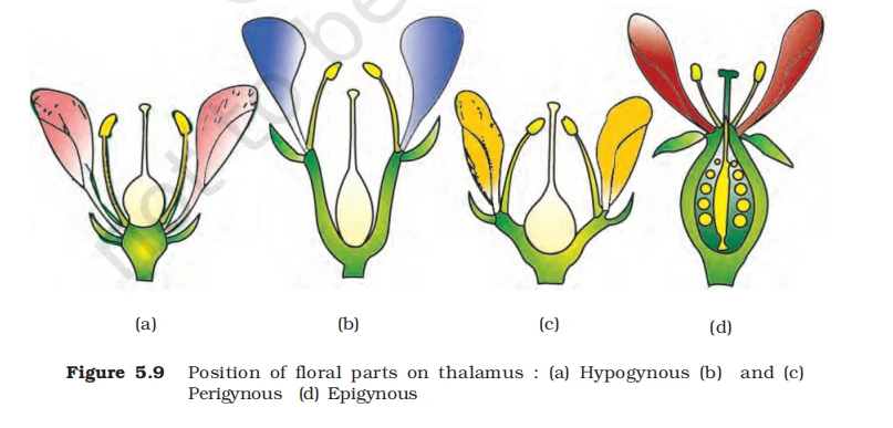
NEET Biology Types of flowers based on the position of ovary
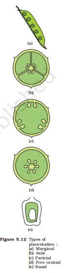
NEET Biology Types of placentation
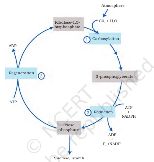
NEET Biology C3 cycle
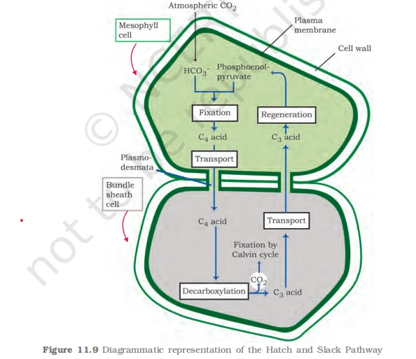
NEET Biology C4
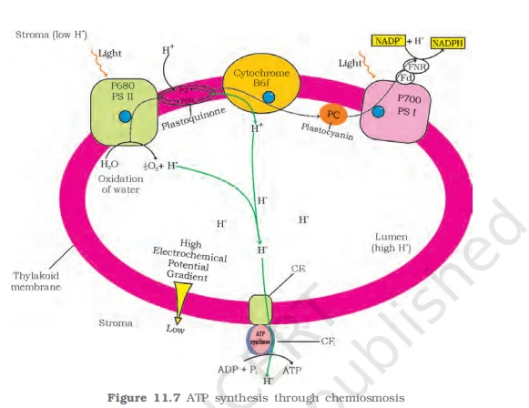
NEET Biology Chemiosmosis
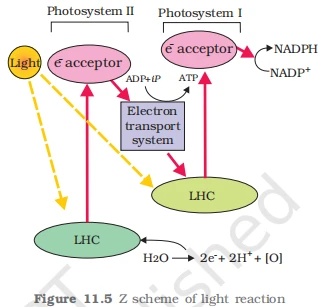
NEET Biology Z scheme of light reaction

NEET Biology Citric acid cycle
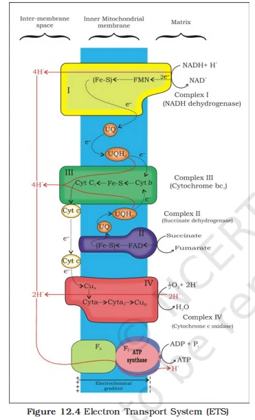
NEET Biology Electron Transport System (ETS)
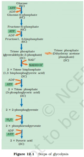
NEET Biology Steps of glycolysis
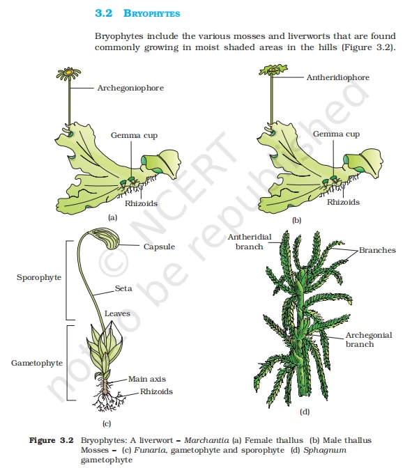
NEET Biology Bryophytes
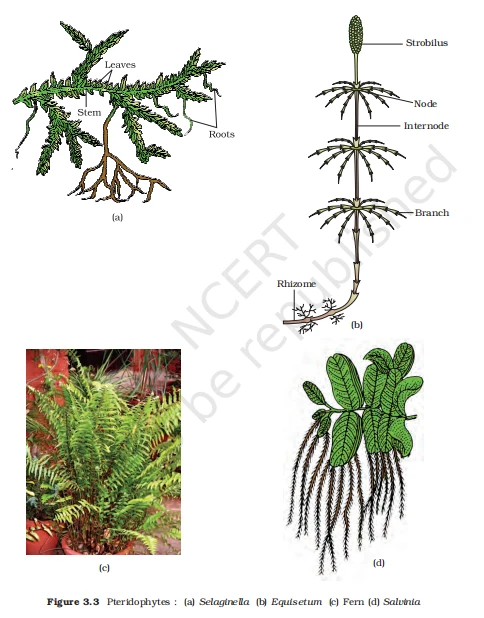
NEET Biology Pteridophytes
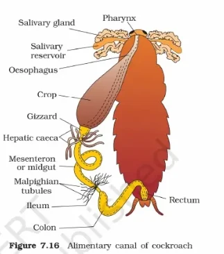
NEET Biology Alimentary Canal of Cockroach
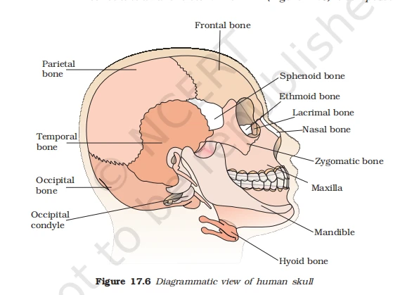
NEET Biology Diagram of human skull
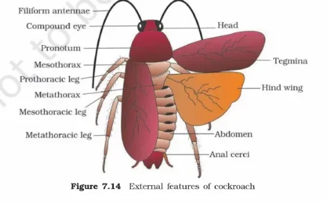
NEET Biology External features of cockroach
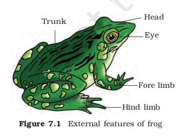
NEET Biology Features of Frog
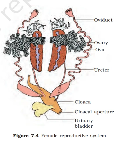
NEET Biology Female reproductive system
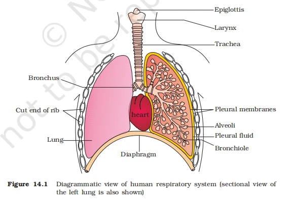
NEET Biology Human Respiratory System
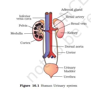
NEET Biology Human Urinary System
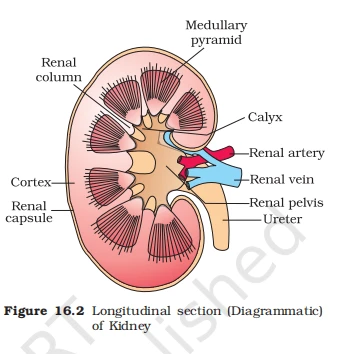
NEET Biology Longitudinal Section of Kidney
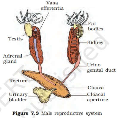
NEET Biology Male reproductive system
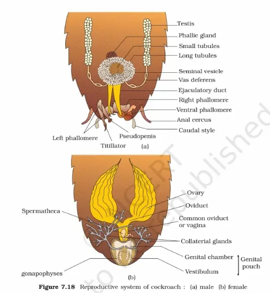
NEET Biology Reproductive system of cockroach
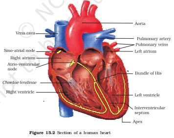
NEET Biology Section of human heart
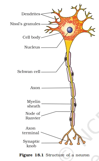
NEET Biology Structure of neuron
Mastered diagrams? Now prove it by solving the NEET 2026 Mock Tests.
Why Diagrams are Important for Class 11 NEET Biology
Diagrams play a vital role in Class 11 NEET Biology preparation for several important reasons:
- Visual Learning: Diagrams for the NEET exam help students visualise complex biological concepts, making them easier to understand and remember. For example, plant kingdom diagrams for NEET improve conceptual clarity.
- Better Memory Retention: Visual aids are easier to recall during exams. A well-practised NEET Biology diagram creates a strong mental image that helps retain information for longer.
- Exam Readiness: The NEET UG exam frequently includes diagram-based questions. The ability to accurately draw and interpret important diagrams for NEET Biology can significantly improve scores.
- Concept Clarity: Class 11 Botany diagrams for NEET break down complicated processes into simple, step-by-step visuals. Topics such as photosynthesis and cell division become much easier to grasp through diagrams.
- Quick Revision: Important diagrams for Class 11 Biology allow fast revision. Instead of rereading lengthy theory, students can quickly review a diagram to recall the complete concept.
Tips to Memorise Important Diagrams in Class 11 Botany for NEET
You should consistently practise important diagrams for Class 11 Botany to strengthen your NEET Biology preparation. Use these proven techniques:
- Use NCERT Textbooks: Start with NCERT Biology Class 11, as it contains the most important diagrams for NEET. Understand every figure along with its labels.
- Create a Diagram Checklist: Prepare a chapter-wise list of important diagrams for NEET Biology to track your progress.
- Practise Drawing Regularly: Repeated practice of Class 11 Botany diagrams improves recall and helps you reproduce neat diagrams in the NEET exam.
- Label Accurately: Always label every part correctly in NEET Biology diagrams. Even small labelling mistakes can cost marks.
- Use Colour Coding: Highlight different parts using colours. This improves visual memory and makes NEET revision more effective.
- Refer to Multiple Sources: Use trusted reference books and quality online resources to strengthen your understanding of NEET important diagrams.
- Try Group Study: Studying with peers helps reinforce NEET Biology concepts. Quiz each other on diagrams and their functions.
- Use Flashcards: Create flashcards featuring important NEET Biology diagrams on one side and labels on the other for quick self-testing.
By applying these strategies, students can effectively master important diagrams for Class 11 Botany and boost their NEET UG Biology score. Make sure to cover the complete NEET Biology syllabus, including both Botany and Zoology, for the best results.